3-D Cone Beam Imaging (CBCT)
Visit Us for Precise Dentistry
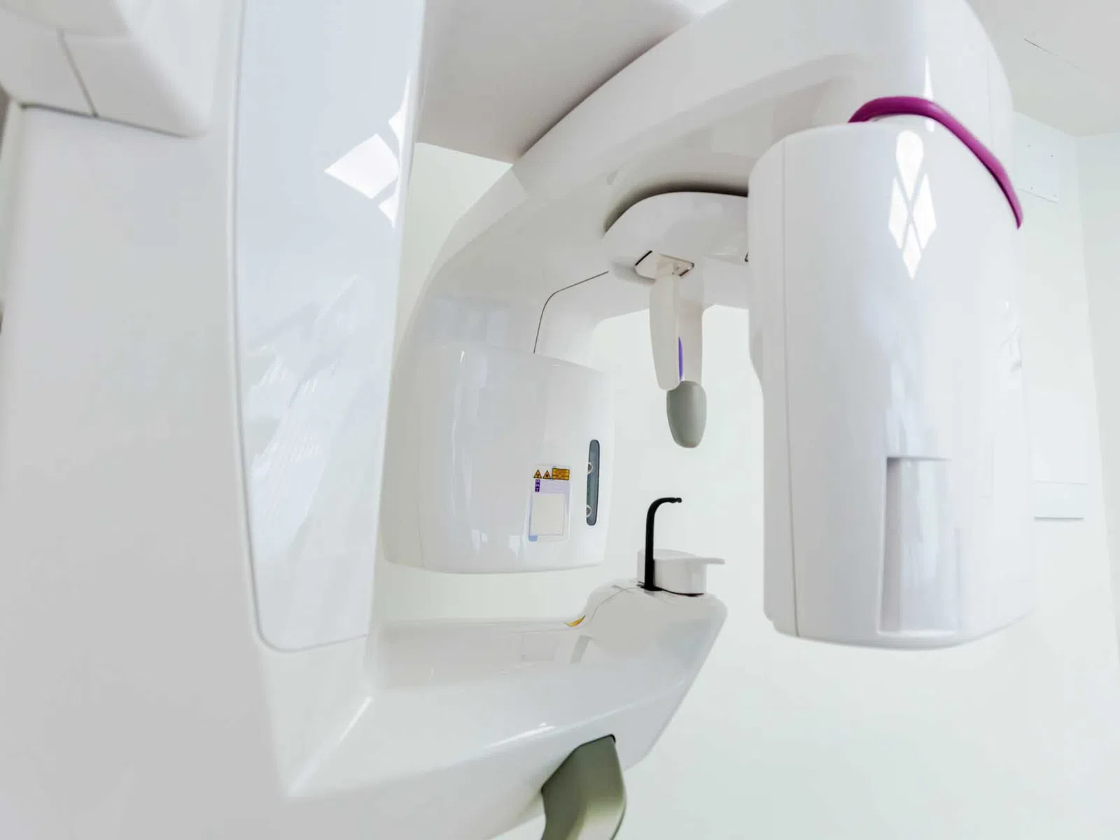
See Beyond the Surface With 3-D Cone Beam Imaging in Knoxville
3-D cone beam imaging in Knoxville provides detailed, three-dimensional views of your teeth, jaws, bone, and surrounding oral structures. At Law Family & Cosmetic Dentistry, CBCT imaging can give your dentist information that isn't always available through a visual examination or conventional two-dimensional dental images.
A CBCT scan captures multiple images from different angles and combines them into a three-dimensional representation of the area being examined. Your dentist can then study anatomical relationships from multiple perspectives, helping provide a clearer understanding of what's happening beneath the surface.
Get a more complete picture when your diagnosis or treatment calls for additional detail.
Learn How Three-Dimensional Dental Imaging Can Improve Your Results
Not every dental concern requires a CBCT scan. When three-dimensional information would be valuable, however, cone beam imaging can reveal details about teeth, roots, jawbone, and nearby anatomical structures. This information can guide diagnosis or treatment planning. The scanning process itself is relatively quick. The imaging unit rotates around your head while capturing the information needed to construct the 3-D image. Unlike a traditional dental X-ray that produces a flat, two-dimensional view, CBCT data can be examined from different angles and perspectives.
That added dimension is particularly valuable when planning treatment in areas where multiple structures are close together. Your dentist may need to evaluate available bone, understand the position of a tooth or root, examine jaw anatomy, or gather additional information before determining an appropriate treatment approach.
When you visit us for 3-D cone beam imaging in Knoxville, you'll benefit from detailed diagnostic information that can support more informed dental care.
Dental Implants
Evaluate available jawbone and nearby anatomical structures by gathering three-dimensional information for dental implant treatment planning.
TMJ Treatment
Examine the bony structures of the jaw joints with three-dimensional imaging as one part of a comprehensive evaluation for TMJ-related concerns.
Oral Surgery
Gain additional information about teeth, roots, bone, and surrounding anatomy when detailed imaging is needed before certain surgical procedures.
Full-Mouth Reconstruction
Examine teeth, bone, and supporting structures from multiple perspectives when coordinating complex restorative care involving several areas of the smile.
Dental Emergencies
Gather additional diagnostic information in select situations when dental pain, trauma, or another urgent concern requires a more detailed view of the affected area.
Frequently Asked Questions About 3-D Cone Beam Imaging (CBCT)
Q What is 3-D cone beam imaging?
3-D cone beam imaging is a dental imaging technique that creates three-dimensional views of teeth, jaws, bone, and surrounding anatomical structures. It is also known as cone beam computed tomography, or CBCT.
Q How is a CBCT scan different from a dental X-ray?
A CBCT scan differs from a conventional dental X-ray because it produces three-dimensional information rather than a flat, two-dimensional image. This allows dental professionals to examine certain anatomical structures from multiple angles when additional detail is needed.
Q What can a CBCT scan show?
A CBCT scan can show detailed information about teeth, tooth roots, jawbone, and surrounding anatomical structures. What a dentist evaluates on the scan depends on the condition being investigated or the treatment being planned.
Q Why is CBCT used for dental implants?
CBCT may be used for dental implants because three-dimensional imaging can provide information about available jawbone and nearby anatomical structures. This information can help a dentist evaluate important factors involved in implant treatment planning.
Q Can CBCT imaging be used before oral surgery?
CBCT imaging can be used before certain oral surgery procedures when a three-dimensional view of teeth, roots, bone, or surrounding anatomy would provide useful information. Whether a scan is necessary depends on the individual procedure and patient.
Q Can CBCT imaging help evaluate the TMJ?
CBCT imaging can provide detailed views of the bony structures of the temporomandibular joints (TMJs). Because TMJ disorders can involve muscles, soft tissues, the bite, and other factors, CBCT is only one potential component of a broader diagnostic evaluation.
Q Does everyone need a CBCT scan?
Everyone does not need a CBCT scan. Three-dimensional imaging is generally recommended when the additional information it provides is useful for diagnosis, treatment planning, or evaluation of a particular dental concern.
Q
How long does a dental CBCT scan take?
A dental CBCT scan generally captures the necessary images in a relatively short amount of time, although the exact scanning process varies by system and the area being examined. Patients typically remain still while the imaging unit rotates around the head to collect the data needed for the three-dimensional image.